Shining a Light on Zonular Microfibrils
Even before joining the University of Health Sciences and Pharmacy community, Juan Rodriguez, Ph.D., professor of physics, carried with him an interest in ophthalmology research. Taking advantage of the University’s unique location on the Washington University Medical Campus, Rodriguez began to attend ophthalmology seminars at Washington University School of Medicine in St. Louis, where he connected with Steven Bassnett, Ph.D., Grace Nelson Lacy Distinguished Professor of Opthalmology and Visual Science at the School of Medicine.
Rodriguez and Bassnett are now collaborating on research investigating the elasticity of the zonular fibers found in the eye, specifically those that help suspend the lens within the eye and are located directly behind the iris.
Imagine a trampoline with elastic ropes holding the center of the trampoline in place — that is essentially what these fibers do. Like a scaffolding, connective tissues, like the zonular fibers we are studying, hold other tissues together and provide those tissues with elasticity.
Rodriguez was recently awarded a grant from the Ines Mandl Research Foundation, which will allow him to take a sabbatical and focus solely on his research.
“The purpose of the grant is to investigate what makes these fibers elastic, beginning with the proteins called fibrillin that make up the fibers, and how those proteins allow these fibers to last for a lifetime, as they contract and expand within the eye,” Rodriguez said. “Unlike other fibers in the body, the fibers found in the eye are fully exposed and are only surrounded by fluid, making them very accessible. The problem is that we are using zonular fibers from mice, and the fibers are very tiny with a width about 200 times thinner than a human hair, and those fibers are made of even smaller fibers.”
To study and capture images of these fibers through light deflection, Rodriguez uses an Atomic Force Microscope (AFM) housed in the Alafi Neuroimaging Laboratory on the Washington University Medical Campus. The AFM is able to capture images and measure samples down to a scale of a million times smaller than the width of a human hair.
However, Rodriguez is still fine-tuning how best to retrieve effective samples of the fibers — approaches he is exploring in the physics labs of UHSP.
“You have to grab both ends and then somehow break it free without damaging it,” Rodriguez explained. “I may have found a way to grab both ends but breaking it free is a challenge. In the physics lab the other day, I was working with a new tool, a laser engraver and cutter. The tool involves a laser that goes around and creates patterns, or when the beam is very intense, it can actually cut thin pieces of material. That got me thinking that maybe I could cut them with a laser to release the fibers.”
The samples that Rodriguez has been able to look at under the AFM have already yielded some unexpected observations. When reviewing the most recent images of the microfibrils, Rodriguez noticed something unusual about the structure of these particular fibers.
“The ‘beads’ are missing — typically there would be an image of a bead about every 2 millionths of an inch along microfibrils, or at least structurally that is what we expect,” Rodriguez explained. “That was something unexpected. It could be just an artifact, or it may be an important clue to how microfibrils transform when bundled into larger fibers. It is definitely something we will have to investigate.”
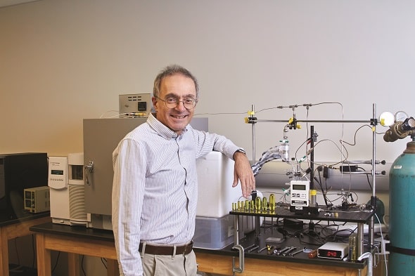
Though Rodriguez has already spent plenty of time in the Alafi Neuroimaging Laboratory, he spends the majority of his time in the physics lab housed in Jones Hall on the UHSP campus.
“The physics lab on campus is partly dedicated to teaching, but there also is a whole area devoted to research,” Rodriguez said. “I plan to continue developing several prototypes on campus to later test out in the School of Medicine’s lab.”
One of the things I hope comes from this research project is that it demonstrates to students how relevant physics is to biology and medicine — most biomedical research today doesn’t take place in just a single lab. It involves the collaboration of many researchers across many disciplines.
This story was first published in the fall 2022 issue of Script Magazine. To view past issues of Script, visit the Script Magazine archive.


